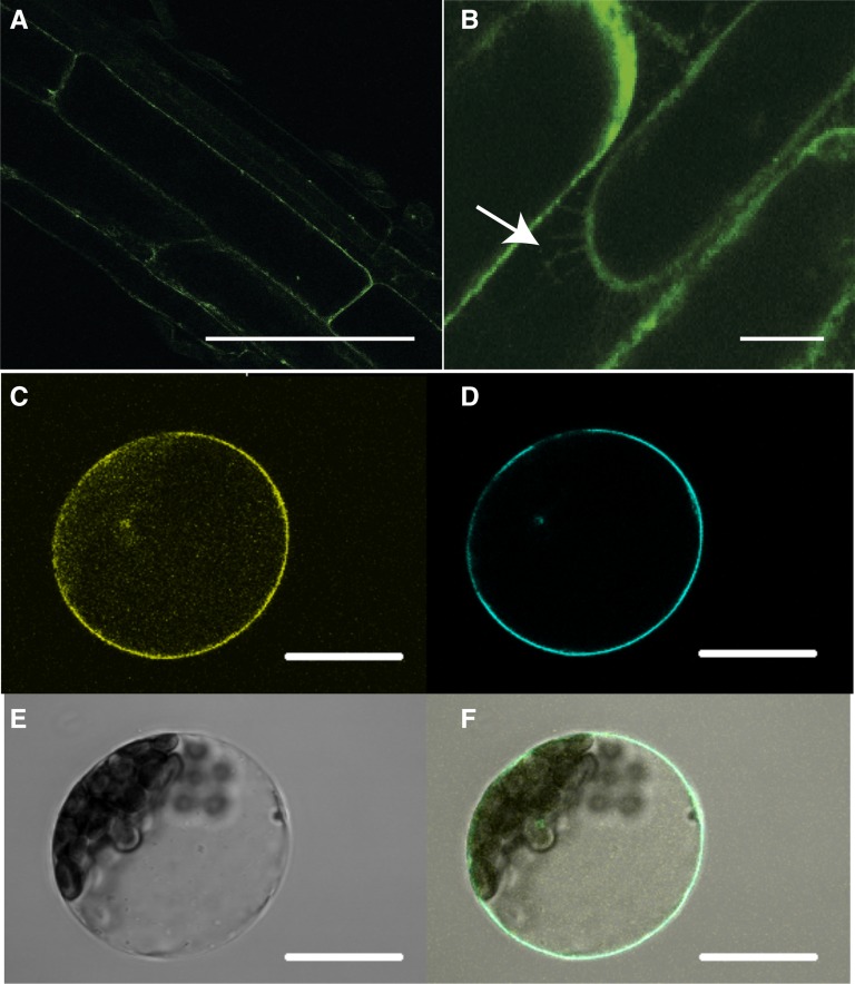
Figure 6.
NPF2.4 protein localizes to the plasma membrane. A and B, Constitutive stable expression of GFP::NPF2.4 in the Arabidopsis root. Bars = 20 µm. A, Confocal image of root cells of 10-d-old GFP::NPF2.4-expressing plants showing the plasma membrane localization of GFP tagged NPF2.4. B, Plasmolysis performed on the GFP::NPF2.4-expressing plants showing Hechtian strands as indicated by the white arrows. C to E, Transient expression of YFP::NPF2.4 fusion construct in Arabidopsis mesophyll protoplasts. Bars = 100 µm. C, Confocal image of YFP::NPF2.4 fusion protein in Arabidopsis mesophyll protoplasts. D, Cyan fluorescence of plasma membrane marker ECFP::ROP11. E, Bright-field image of the transformed protoplast. F, Merged image showing the colocalization of YFP::NPF2.4 fusion protein and the plasma membrane marker.

