Figure 4.
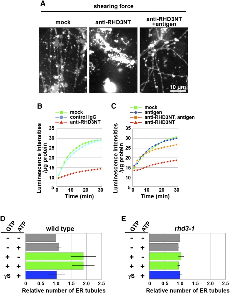
In vitro requirement of RHD3 for membrane fusion and subsequent tubule formation. A, An ER vesicle preparation from cultured Arabidopsis MM2d cells was preincubated with each of buffer solution (mock) and anti-RHD3NT (no antigen) and preadsorbed anti-RHD3NT with the antigen peptide (2.5 µm, antigen) and was then subjected to the in vitro ER tubule elongation assay under a shearing force. Note that anti-RHD3NT abolished ER tubule formation. B and C, Ca2+-efflux-based assay. ER vesicles prepared from cultured Arabidopsis MM2d cells were preincubated with each of buffer solution (mock), control IgG, and anti-RHD3NT (B), and another preparation was preincubated with each of buffer solution (mock), the antigen peptide, anti-RHD3NT, and preadsorbed anti-RHD3NT with the antigen peptide (C). Luminescent intensities per microgram of protein were plotted against time after addition of ATP and GTP. Note that anti-RHD3NT abolished the GTP-dependent enhancement of luminescent intensity. See Supplemental Table S3 for statistical analyses of increasing intensity in each condition. D and E, Quantitative analysis of in vitro ER tubule formation from ER vesicles prepared from wild-type (D) and rhd3-1 (E) seedlings that expressed ER-luminal GFP. ER vesicle preparations that were treated with nucleotides were subjected to an in vitro ER tubule elongation assay under the shearing force. The x axis indicates the number of ER tubules formed relative to the number ER tubules formed in the absence of the nucleotides. The experiments were repeated at least three times. Error bars indicate sd. See Supplemental Table S4 for statistical analyses of the number of ER tubule in each condition.

