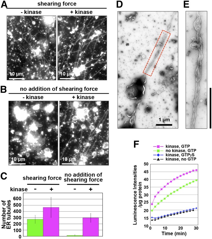
Figure 6.
Kinase-dependent enhancement of ER membrane fusion and ER tubule formation activities. A to C, In vitro ER tubule formation from the kinase-treated and untreated ER vesicles with (A) or without (B) an applied shearing force. The assay is described in Figure 3E. In B, to minimize shearing forces during the assay, the specimen was prepared on ice. Note that kinase treatment enabled the ER vesicles to form ER tubules without an applied shearing force. C, Number of the ER tubules under the condition described in A and B was counted. D and E, Negative-staining electron micrographs of an ER tubule emerging from a kinase-treated ER vesicle. Boxed region in D was enlarged in E. Bars = 1 µm. F, Ca2+-efflux-based assay of an ER vesicle preparation from cultured Arabidopsis MM2d cells. An ER vesicle preparation was treated with kinase in the presence of GTP or a nonhydrolyzable GTP analog (GTPγS). The calcium ion efflux produced by the ER vesicles was monitored by aequorin. Luminescent intensities per microgram of protein were plotted against time after addition of ATP and GTP. Note that luminescent intensity was increased by adding GTP.

