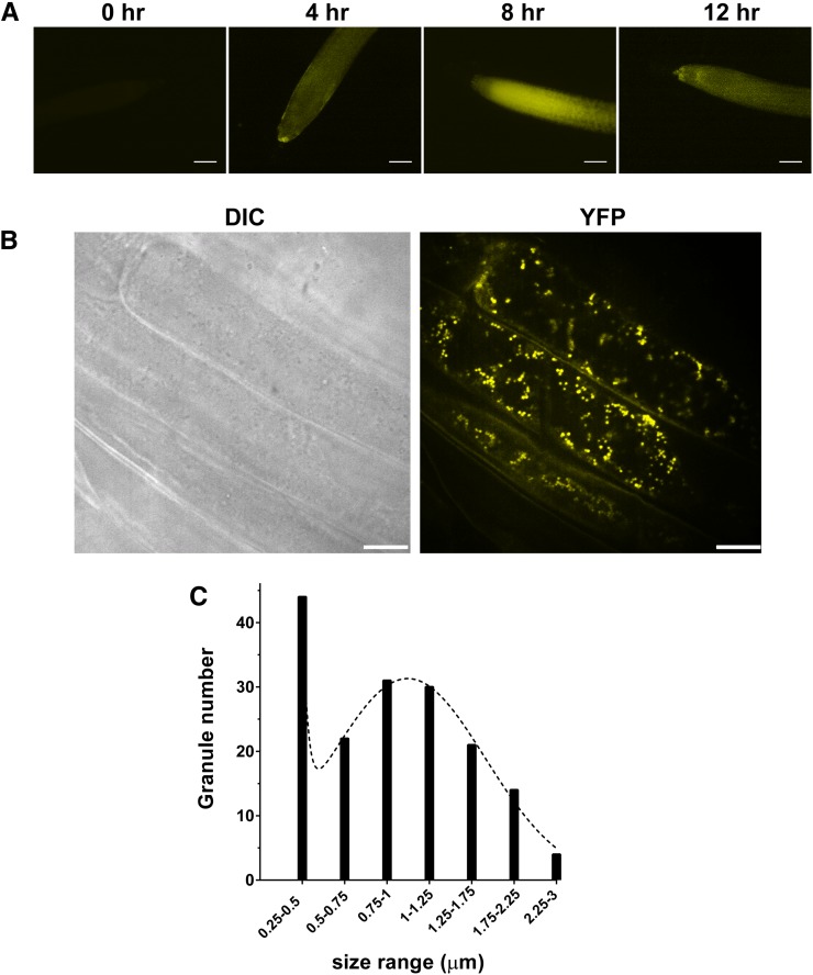
Figure 4.
Subcellular localization of CML38-YFP in the roots of hypoxia-challenged 10-d-old CML38-YFP recombineering seedlings. A, Ten-day-old vertically grown CML38-YFP seedlings were subjected to anaerobic stress by complete submergence in water, and the appearance CML38-YFP protein was monitored by fluorescence microscopy. B, Higher resolution confocal micrographs of root cells from seedlings after 8 h of submergence. DIC represents the image obtained with differential interference contrast optics, while YFP represents the image obtained with the YFP filter set. C, Histogram of the size distribution of CML38-YFP granules (between 0.4 and 3 μm) from two representative cells as determined by ImageJ analysis. The dotted line shows the fit to a sum of two Gaussian distributions. The median particle size was 0.9 μm, and the average was 1 μm. Bars = 100 µm (A) and 25 µm (B).

