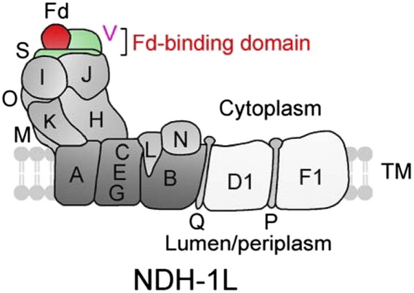
Figure 8.
A model schematically represents the localization of NdhV in Fd-binding domain of cyanobacterial NDH-1 complex. NdhV, which was suggested to be located at the surface of NdhS, may stabilize the binding of NdhS with reduced Fd in Fd-binding domain of cyanobacterial NDH-1 complex. NdhS and NdhV are two OPS subunits and are indicated by green. TM, Thylakoid membrane.

