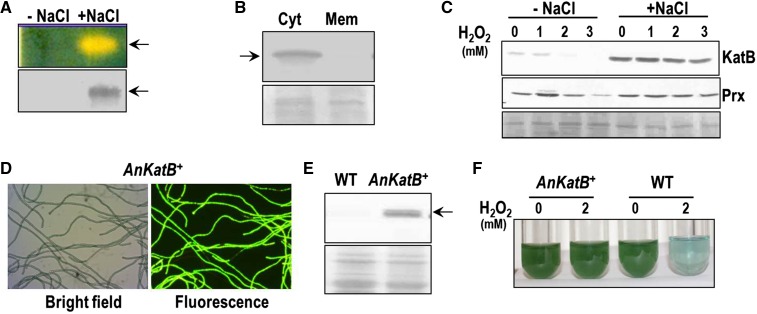
Figure 2.
Expression of KatB in Anabaena PCC 7120. A, The cell free extracts of control (-NaCl) and NaCl-treated (+NaCl) Anabaena PCC 7120 were assayed for in gel catalase activity (zymogram, top) or probed with the KatB antiserum at 1:10,000 dilution on a western blot (bottom). The experiment was repeated thrice with similar results and a representative image is shown. B, Localization of KatB. The salt-stressed Anabaena cells were lysed with glass beads, and the membrane proteins in the lysate were separated from the cytosolic proteins. Cytosolic and membrane proteins were resolved on SDS-PAGE, transferred onto a nitrocellulose membrane, and probed with the KatB antiserum. The Ponceau S-stained part of the nitrocellulose membrane is shown as a loading control at the bottom. C, The cell-free extracts of control (-NaCl) or NaCl-pretreated (+NaCl) Anabaena exposed to H2O2 were resolved on SDS-PAGE and electroblotted on to nitrocellulose membrane. These were probed with the KatB antiserum (top, KatB) or the Alr4641 (2-Cys-Prx) antiserum (bottom, Prx). The Ponceau S-stained part of the nitrocellulose membrane is shown as a loading control at the bottom. D, Bright field and fluorescence micrograph of KatB overexpressing Anabaena strain (AnKatB+). E, Overexpression of KatB protein in Anabaena. The cell-free extracts of the wild-type Anabaena PCC 7120 (WT) and AnKatB+ were resolved by SDS-PAGE, immunoblotted, and probed with anti-KatB antiserum. Ponceau S-stained loading control is shown at the bottom. F, The wild-type Anabaena PCC 7120 (WT) or Anabaena overexpressing KatB (AnKatB+) cells were treated with H2O2 (2 mm) for 2 d and photographed.

