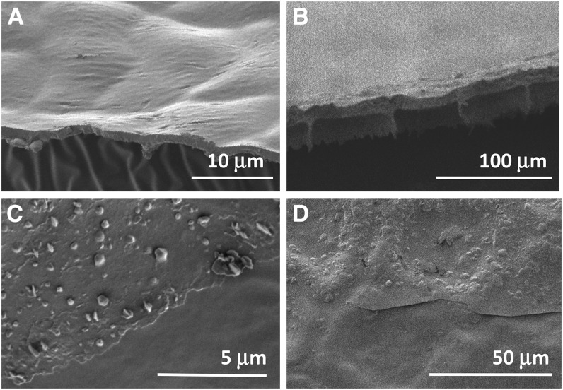
Figure 1.
Scanning electron micrographs of isolated cuticles. A and B, Side views of cryofractured cuticles from leaves of Citrus aurantium (A) and M. deliciosa (B), illustrating species with relatively thin and thick cuticles, respectively. C and D, Top-down views of the outer surface of cuticles of Citrus aurantium (C) and M. deliciosa (D). The bottom portions of the images show areas from which the epicuticular wax film was removed with adhesive, while the top portions show the native cuticle surface.

