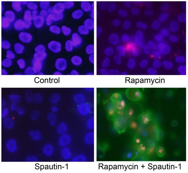
Figure 6.
Fluorescent staining for the detection of apoptosis. Apoptotic cells were detected by an Annexin V/propidium iodide (PI) and Hoechst 33342 triple-staining assay. Cells were stained by blue (Hoechst 33342), green (Annexin V) and red (PI). There were more Annexin V-stained cells in the group treated with both rapamycin and Spautin-1 than in the other groups. Rap, rapamycin; Spa, Spautin-1.

