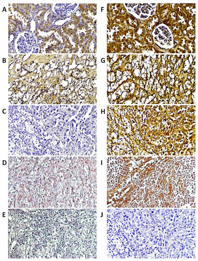
Figure 6.
Immuhistochemical localization of RASSF1A and RASSF1C proteins in ccRCC. Immuhistochemical localization of (A–D) RASSF1A and RASSF1C (F–I) proteins in ccRCC. Normal kidney (A and F), tumor kidney of TNM 3 and Fuhrman's grade 2 (B and G) or TNM 4, Fuhrman's grade 4 (C and H) or metastasized lymph node (D and I) of two ccRCC patients (according to Fig. 5) are shown. Strong reaction was observed for RASSF1C in all samples, RASSF1A is characterized by strong presence in epithelial cells of control kidney; weak expression was observed in tumor and mestastized samples as compared to negative control (primary antibody was omitted) of (J) either tumor or (E) metastasized lymph node samples. Magnification, ×20.

