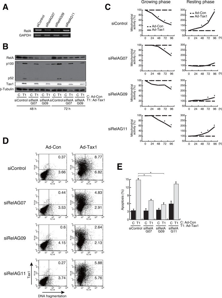
Fig 7. Participation of RelA in Tax1-mediated growth inhibition and apoptosis.
(A) Growing Kit 225 cells were transfected with RelA-specific siRNA and cultured for 72 h. RelA expression was examined by RT-PCR. GAPDH was used as an internal control. (B) Growing Kit 225 cells were transfected with RelA-specific siRNA 24 h before adenovirus infection (Ad-Tax1 or Ad-Con), and harvested 48 h and 72 h post infection for western blotting. RelA, p100, p52 and Tax1 expression was monitored by immunoblotting with anti-RelA, anti-p52 and anti-Tax1 antibodies. β-Tubulin was used as an internal control. (C) siRNA-treated growing or resting Kit 225 cells were infected with Ad-Tax1 or Ad-Con, and cultured for indicated times. Mitochondrial activity was measured by MTT assay. Relative percentages of Ad-Tax1 to Ad-Con are shown. *, p<0.05. (D) siRNA-treated growing Kit 225 cells were infected with Ad-Tax1 or Ad-Con, and cultured for 72 h. Tax1 expression and DNA fragmentation were measured by flow cytometry. (E) Percentage average number of cells undergoing apoptosis was calculated from three independent experiments. Values are shown as the means ± SE. *, p < 0.05.

