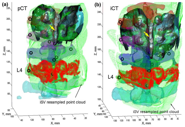
Fig. 6.
Individually segmented and color-coded vertebrae in pCT (a) and iCT (b) overlaid with the uniformly resampled iSV point cloud as well as the implanted mini-screws (black circles). A typical reconstructed iSV texture surface is also shown. For illustration, the iSV point clouds corresponding to L4 are highlighted

