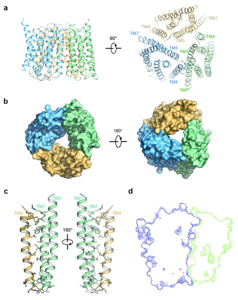
Figure 3. Structure of the OsSWEET2b trimer.
a, Two orthogonal views of the OsSWEET2b trimer in ribbon representation. b, Surface representations of OsSWEET2b viewed from the intrafacial (cytosolic; left) and extrafacial (luminal; right) side. c, Close-up view of the trimer interface. TM4 from one protomer and TM5 and TM7 from the neighbouring protomer are shown as ribbon representation. Side chains of residues participating in the interaction are shown as sticks. d, Cross-section view of the trimer interface. The two interacting protomers are presented as blue and green surface meshes.

