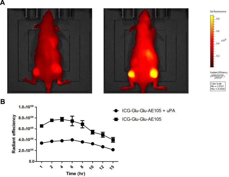
Fig 5. In vivo blocking of ICG-Glu-Glu-AE105 by uPA, the natural ligand.
(A) Representative images obtained by the IVIS Lumina XR at 710 nm showing a mouse receiving uPA simultaneously with ICG-Glu-Glu-AE105 resulting in decreased signal compared to a mouse only receiving ICG-Glu-Glu-AE105. (B) Two groups of mice (n = 4) were dynamically scanned with either ICG-Glu-Glu-AE105 + uPA or ICG-Glu-Glu-AE105. In all timepoints the two groups were significantly different with the group receiving only ICG-Glu-Glu-E105 having a 2 fold higher signal.

