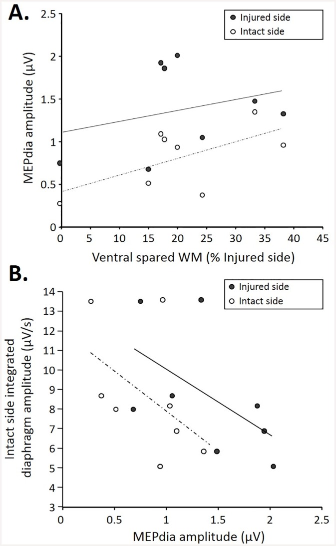
Fig 4. Correlations among MEPdia amplitude, extent of injury, and contralateral diaphragm activity at 7d P.I.

A. Correlation of the extent of injury (represented as the ventral spared white matter) with MEPdia amplitude on the injured (black dots, R2:0.16, p = 0.034) and spared (white dots, R2:0.364, p = 0.149) side. Note the higher MEPdia recording with bigger spared white matter. B. Correlation between contralateral diaphragm amplitude and MEPdia amplitude on injured (black dots, R2:0.559, p = 0.005)/spared (white dots, R2:0.495, p = 0.014)sides. Note higher diaphragm EMG with lower MEPdia amplitude.
