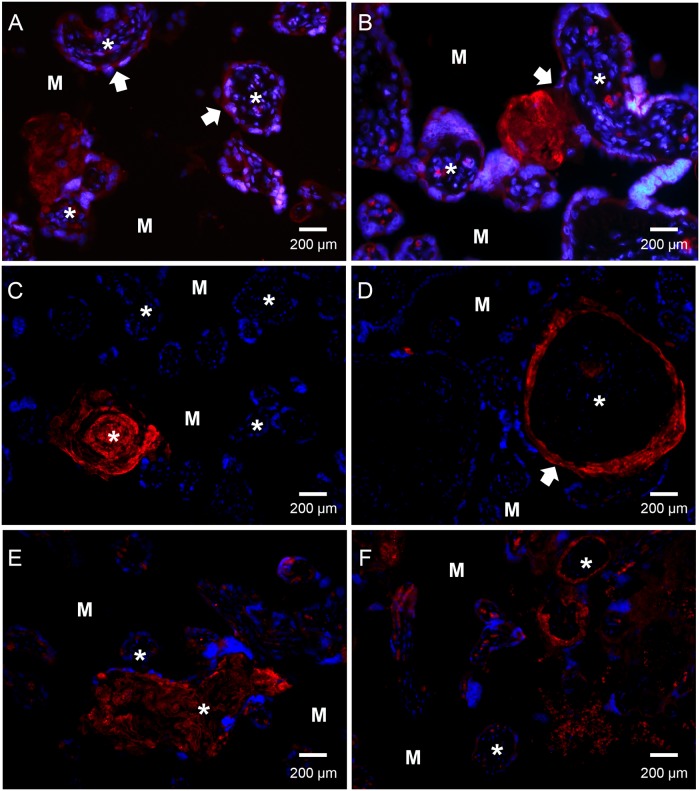
Fig 2. Immunofluorescence microscopy detection of SALSA localization in human placenta.
Paraffin-embedded tissue from healthy (panels A-D) and pre-eclamptic (PE) (panels E-F) placentas. Sections were stained with an anti-SALSA antibody (Hyb 213–06) and Alexa 546-conjugated goat anti-mouse IgG. Red: SALSA, blue: DAPI. * denotes the center of the villus structures. M denotes the intervillous space/maternal tissue. Panels A to F show a widespread but focal staining of SALSA in the human placenta. Based on the morphology and localization of the placental structures SALSA appears to be present in fibrinoid formations, especially in relation to a disruption of the syncytiotrophoblast layer. (A) shows the expression of SALSA found intracellularly in the syncytiotrophoblast layer of some, but not all, villi (white arrowhead). SALSA was observed abundantly in fibrinoid structures at the edge of the villi. A breach of the syncytiotrophoblast layer accompanied by formation of SALSA-positive fibrinoid in the intervillous space can be observed (white arrowhead, panel B). (C) shows SALSA in a necrotic villus with fibrinoid formation. (D) shows a villus with disrupted syncytiotrophoblast layer (white arrowhead). Here the disruption may have led to the influx of maternal blood, and SALSA-positive fibrinoid can be observed separating the syncytiotrophoblast layer from the underlying basement membrane. The fibrinoid may deposit all the way around the exposed villus structure, thus forming a ring structure. Syncytial damage and fibrinoid formation is observed more frequently in PE. (E) and (F) show examples of a necrotic villus and a ring formation in PE placentas. 200× magnifications.

