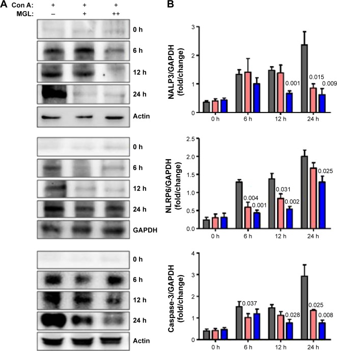
Figure 8.
The inflammasome expression in liver tissue.
Notes: After Con A challenge for 6, 12, and 24 hours, the NALP3, NLRP6, and caspase-3 expression increased. (A) Shows the results of the Western blotting analysis, and (B) shows the plots of pixel intensity. For simplicity, representative blots of actin and GAPDH are shown. MGL administration significantly downregulated inflammasome expression, in which caspase-3 decreased in a dose-dependent manner.
Abbreviations: Con A, concanavalin A; MGL, magnesium isoglycyrrhizinate.

