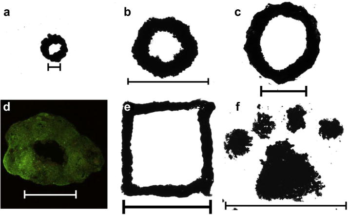
Fig. 4.

JMCSs aligned and fused into homogenous tissues using magnetic patterning. (a–c) JMCSs composed of primary rat aortic smooth muscle cells were patterned and fused together into rings of varying sizes, ranging from 2 mm (a, 25 JMCSs, scale bar = 1000 μm) up to 10 mm (b, c, 3000 JMCSs each, scale bar = 10 mm). (d) Furthermore, viability of the fused structure was confirmed using simultaneous live/dead (green/red) fluorescent staining (scale bar = 1000 μm). (e, f) Finally, using various magnetic patterns, spheroids assembled onto custom patterns can fuse together over the course of days to form fused constructs, demonstrated by a square (e) and Clemson University Tiger Paw (f). Samples shown in images were allowed to fuse over the course of 4 days (scale bars: e = 5 mm, f = 10 mm). (For interpretation of the references to color in this figure legend, the reader is referred to the web version of this article.)
