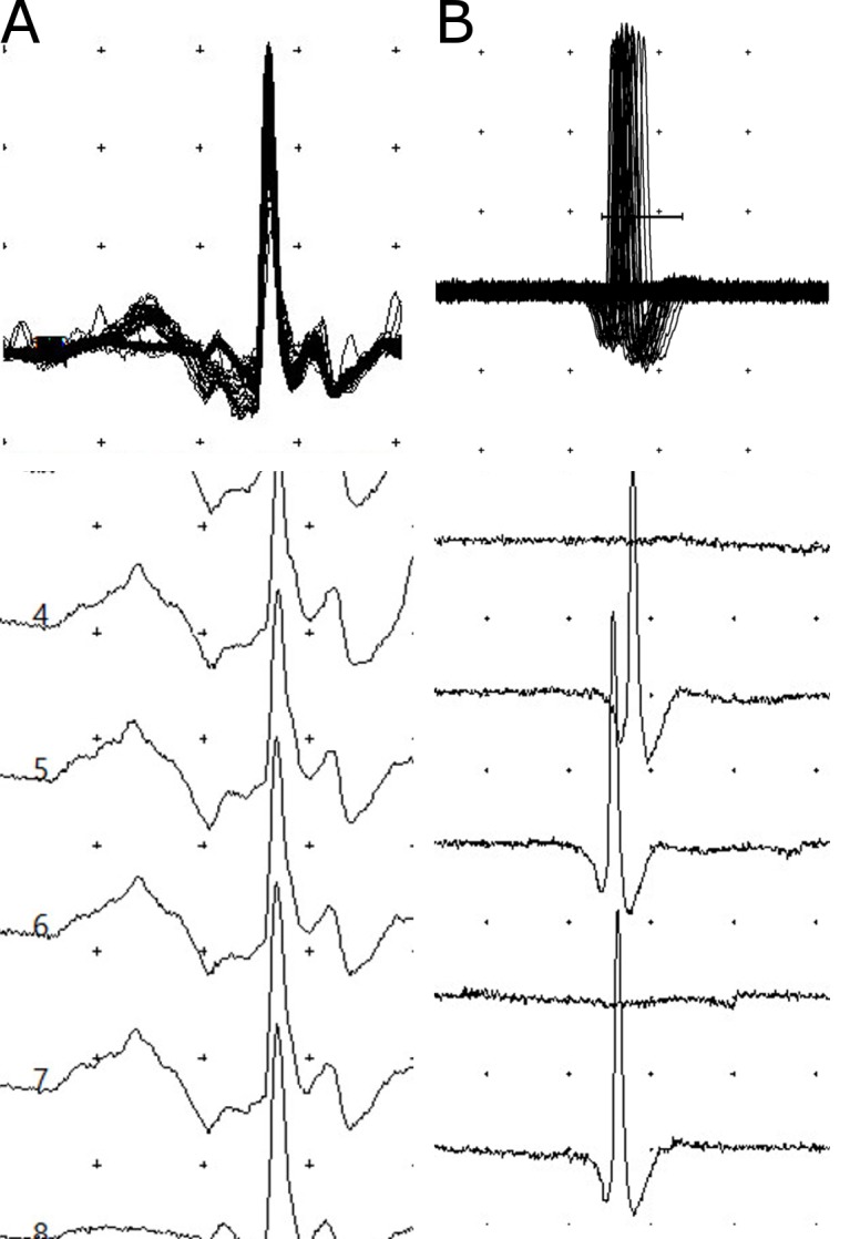
Fig 3. Superimposed and non-superimposed sfEMG recordings of the orbicularis oculi muscle of patients following krait bites.

A, recordings of a patient on admission with no neurotoxicity indicating the normal jitter (14.5μs) and no blocks; B, high jitter (61.6μs) with intermittent blocks seen in a patient on admission with severe neuromuscular paralysis. (The distance between two dots represents 200μV vertically and 3ms horizontally.)
