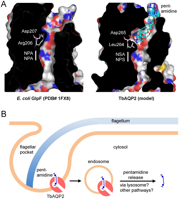
Fig 4. Model of the pentamidine binding mode to TbAQP2 and proposed uptake by endocytosis in the flagellar pocket.
(A) Shown are the crystal structure of the prototypical aquaglyceroporin GlpF and a model of TbAQP2. GlpF Arg206 and TbAQP2 Leu264 mark the position of the ar/R selectivity filter. In TbAQP2, the Asp265 sidechain carboxylate binds to an amidine moiety of pentamidine (light blue), whereas in GlpF the space is occupied by the guanidine sidechain of Arg206. The location of the ‘NPA/NPA’ region (white bar) and sequence deviations in TbAQP2 are indicated. (B) Proposed uptake mechanism of pentamidine via high-affinity binding to TbAQP2, endocytosis of the complex, and release of pentamidine in the acidic lysosome due to pH shift or TbAQP2 degradation.

