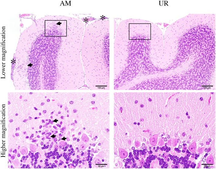
Fig 9. Inflammatory infiltration in the cerebellum of SAFV-3-inoculated young BALB/c mice.
BALB/c mice were inoculated intracerebrally with 104 CCID50 (cell culture infectious dose) of the aseptic meningitis (AM) and upper respiratory (UR) strains of Saffold virus type 3 (SAFV-3). Hematoxylin and eosin (H&E) staining. Bar, 100 μm (upper panels) and 20 μm (lower panels). Histopathological findings in the cerebellum on Day 8 p.i. Inflammatory cells were observed in the cerebellar cortex (arrows, low magnification) and meninges (asterisk) of AM-inoculated mice (left panels), but not in those of UR-inoculated mice (right panels). Mononuclear cells and rod-shaped cells (arrows, high magnification) were observed in the cortex and meninges of AM-inoculated, but not UR-inoculated, mice (lower panels). Purkinje cells were present in the lesion. The figure shows representative data from two experiments with similar results. Original magnification: upper panels, 200×, and lower panels, 1,000×.

