Figure 1.
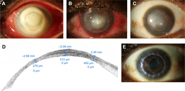
Slit lamp photograph of the right eye of 35-year-old contact lens wearer with Pseudomonas keratitis.
Notes: (A) At presentation, a suppurative infiltrate encompassing most of the cornea as well as a 3.5 mm hypopyon is seen. (B) One month after treatment with fortified topical antibiotics, cornea collagen cross-linking, amniotic membrane transplantation, and topical corticosteroids, the infiltrate is much smaller and peripheral corneal neovascularization and scarring are observed. (C) Six months later, diffuse scarring limits the patient’s vision. (D) Anterior segment OCT at 1 year shows significant peripheral and central thinning. (E) One year after penetrating keratoplasty, the patient’s vision is 20/60.
Abbreviation: OCT, optical coherence tomography.

