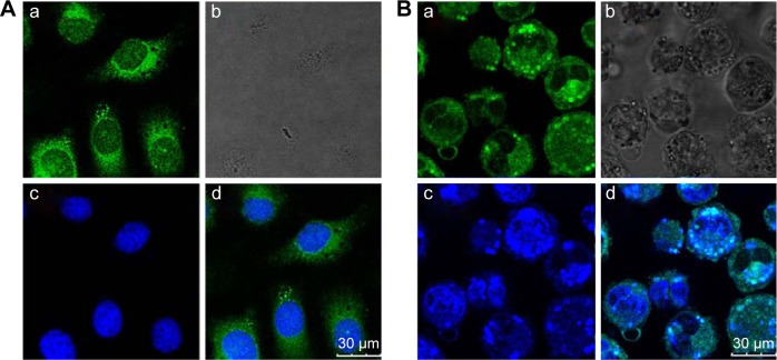
Figure 3.
Confocal laser scanning microscopy images of HepG2 (A) and HCa-F (B) cells after incubation with 200 μg/mL RCPTN for 4 hours at 37°C.
Notes: The RCPTN were green, while nuclei of the cells were blue stained using DAPI. (A[a]) and (B[a]) are green fluorescence of RCPTN uptaken by HepG2 cells and HCa-F cells, respectively. (A[b]) and (B[b]) are bright field images of HepG2 cells and HCa-F cells, respectively. (A[c]) and (B[c]) are blue fluorescence of HepG2 cells and HCa-F cells nuclei dyed by DAPI. (A[d]) is a combined image of (A[a]) and (A[c]), and (B[d]) is a combined image of (B[a]) and (B[c]), respectively.
Abbreviations: RCPTN, Resibufogenin/coumarin-6-loaded poly(lactic-co-glycolic acid)-d-α-tocopheryl polyethylene glycol 1000 succinate nanoparticle; DAPI, 4,6-diamidino-2-phenylindole dihydrochloride.

