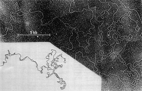
Figure 1. DNA replication bubbles from Drosophila cleavage nuclei.

This figure, reproduced with permission from Kriegstein and Hogness (1974), shows an electron micrograph (and accompanying trace) of DNA purified from very early (< 1 h) fertilised Drosophila melanogaster embryos. This particular molecule shows 23 replication bubbles in a region of 119 kb.
