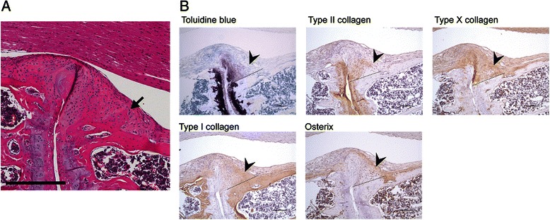
Fig. 4.

New bone and osteophyte formation. a Representative image of an osteophyte (arrow) in the affected joint of a 12-week proteoglycan-induced spondylitis mouse. Scale bar = 300 μm. b Recently formed bone tissue (arrowheads) is characterised by a transition from proteoglycan and type II collagen-enriched matrix to type X and type I collagen-positive matrix. Osterix-positive osteoblasts are embedded in type I collagen-positive bone matrix. The dashed line represents the boundary between the original cortical bone and the excessive matrix above, showing a reduction of cartilaginous matrix, toluidine blue staining and type II collagen accompanied with an increase of type I collagen and osterix-expressing osteoblasts. Arrowheads indicate cartilage tissue that is strongly positive for type II collagen but weakly for type I collagen. Scale bar = 100 μm.
