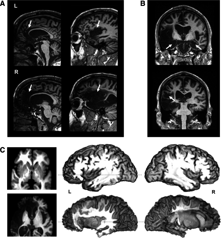
Fig. 1.
Roger’s brain. a Sagittal MRI slices showing bilateral lesions to the ACC (leftmost images) and insula (rightmost images). b Coronal MRI slices showing bilateral lesions to the amygdala (top) and right secondary somatosensory cortex (bottom). c 3D digital “dissection” of the insular cortex: top lateral view of the brain of a healthy non-brain damaged participant, revealing the gyrations of the insular cortex; bottom lateral view of Roger’s brain, highlighting the absence of an insular cortex; left axial MRI slices corresponding to the dashed lines on the 3D-images. All MRI slices are shown in radiological convention. Volumetric analyses (Philippi et al. 2012) reveal that his lesion encompasses 90 % of the insula, 99 % of the ACC, and 100 % of the amygdala. The lesion extends beyond these regions into other limbic territories with more extensive damage in the right hemisphere. The entire right insula is destroyed and the damage in the posterior sector extends into parietal operculum, secondary somatosensory cortex, and the underlying white matter. The vast majority of the left insula is also destroyed with the exception of a small island of tissue in the left dorsal anterior insula that appears to be functionally disconnected from the rest of the brain (Philippi et al. 2012). Although the ACC has been destroyed bilaterally, the more dorsal and posterior aspects of Brodmann area 32 appear to be spared in the left hemisphere; however, this remaining tissue is dorsal to the paracingulate sulcus, and is therefore considered part of the paracingulate cortex (and not the ACC proper). Of note, Roger’s lesion has largely spared the brainstem, thalamus, and primary and secondary somatosensory cortices. The only exception is the aforementioned damage to the right secondary somatosensory cortex, as well as some localized atrophy in the right thalamus and right pons. The reader is referred to Fig. 2 and Feinstein et al. 2010 and Philippi et al. 2012 for additional brain scans and a more detailed account of Roger’s damage

