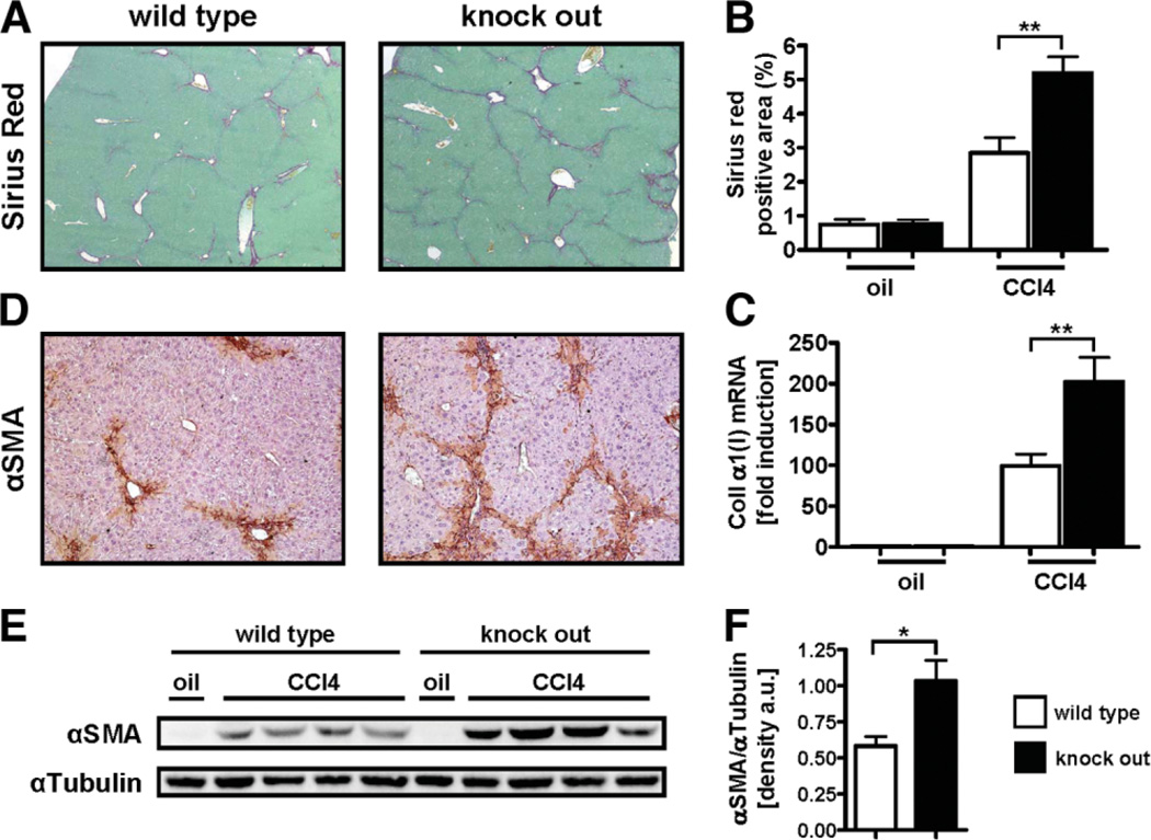
Fig. 3.
Loss of ace2 exacerbates toxic liver injury. Ace2 KO mice (n = 12) and wt littermates (n = 16) received eight injections of CCl4 (0.5 µL/g every 4 days) and were analyzed 48 hours after the last injection. Fibrosis was evaluated by morphometric analysis of Sirius Red-stained sections (A,B), by qPCR for collagen α1(I) (C), by IHC and immunoblotting for αSMA (D,E). Immunoblots were quantified by densitometry (F). Ace2 KO mice displayed significantly increased deposition of fibrillar collagen, mRNA levels of collagen α1(I), and αSMA-positive cells. Data are presented as mean ± SEM; *P < 0.05, **P < 0.01.

