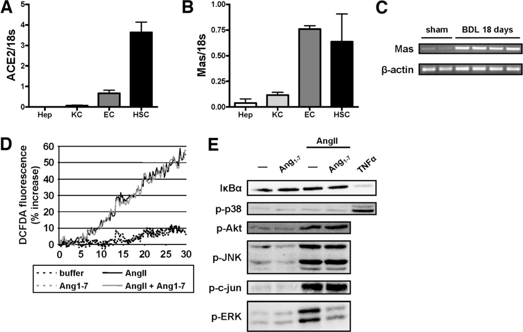
Fig. 7.
Ang1–7 inhibits ERK phosphorylation induced by AngII in HSC. mRNA was isolated from hepatocytes (Hep), KCs, ECs, and HSCs and expression of ace2 and Mas was evaluated by qPCR (A,B). RT-PCR for Mas was performed using mRNA isolated from whole liver from balb/c mice subjected to BDL for 18 days (C). Mouse HSCs were cultured for 5 days and then serum starved for 24 hours. ROS production was evaluated by DCFD fluorescence of mouse HSCs stimulated with AngII (black solid line) and preincubated with Ang1–7 (gray solid line) (D). Cells were stimulated with AngII (10−7 M) in the presence or absence of Ang1–7 (10−7 M) for 15 minutes. Activation of signaling pathways was analyzed by immunoblotting for IκBα and using phospho-specific antibodies for p38, Akt, JNK, c-Jun, and ERK. Equal loading was evaluated by Ponceau S staining (not shown). Data are representative of three independent experiments.

