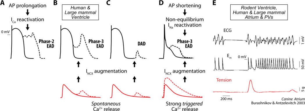
Figure 7. Mechanisms of afterdepolarization formation in cardiomyocytes.
In ventricular myocytes from large mammals, phase-2 EADs are associated with ICa recovery from inactivation and reactivation during prolonged APs (A). Spontaneous SR Ca2+ release, which increases Ca2+ extrusion via NCX (inward current), can lead to phase-3 EADs (B) or DADs (C) when occurring during or after Em repolarization respectively. In murine ventricle, phase-3 EADs are favored by potentiated (triggered) Ca2+ transient and AP shortening (D). The former causes INCX augmentation and AP plateau prolongation (at negative Em), during which non-equilibrium INa reactivation (permitted by rapid INa recovery during fast repolarization) can occur. This, which is shown here to be relevant in the human atrium, could be a universal mechanism underlying EAD formation in both atria (especially PVs) and ventricles ([26]) of large mammals. Indeed, phase-3 EADs mediate re-initiation of atrial (and ventricular) fibrillation. ECG, Em and tension recorded in canine atrium (E, reproduced with permission from [5]) show phase-3 EADs occurring after termination of fast atrial pacing, which are accompanied by increased contractile force.

