
Fig. 1.
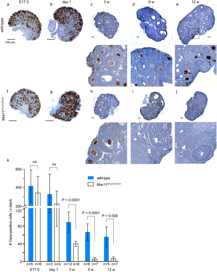
Premature elimination of Mre11 ATLD1/ATLD1 oocytes. a–j Representative images of anti-VASA-stained mid-ovary sections in a–e wild-type and f–j Mre11 ATLD1/ATLD1 ovaries, from E17.5 (a and f), day 1 (b and g), 3-week-old (c and h), 6-week-old (d and i), and 12-week-old (e and j). Bar = 100 μm. k Quantification of the number of follicles. Bars denote the average ± standard deviation (SD). Age and the number of ovaries analyzed were shown below the x-axis. P-value was determined by unpaired t-test. Blue and white bars indicate wild-type and Mre11 ATLD1/ATLD1, respectively