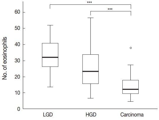
Fig. 1.

The number of eosinophils in the colorectal neoplastic lesions: the number of infiltrating eosinophils increased significantly with the progression of colorectal neoplastic lesions. Asterisk (***) indicates p < .001 in each group. LGD, low grade dysplasia; HGD, high grade dysplasia.
