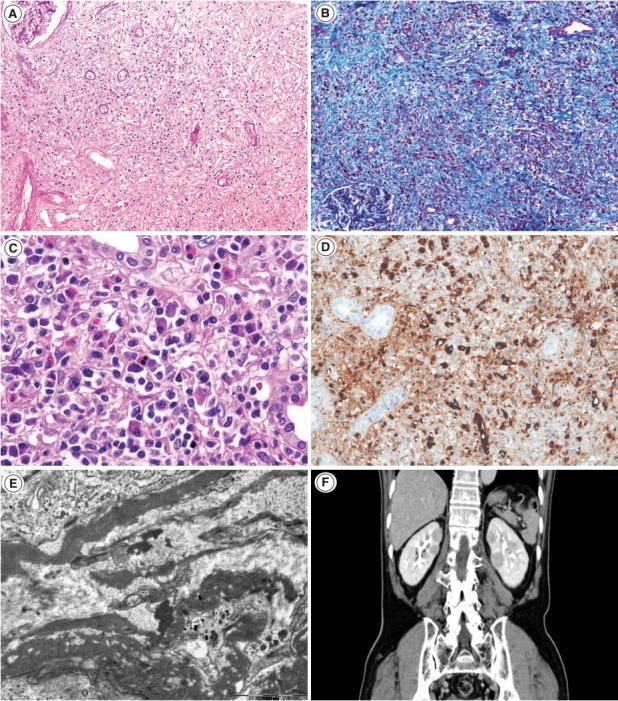
Fig. 1.

Tubulointerstitial nephritis in IgG4-related disease. (A, B) At lower power, interstitial fibrosis is severe and shows a focal streaming pattern with mixed inflammatory infiltration of lymphocytes and plasma cells (A, periodic-acid Schiff ×100; B, Masson trichrome ×100). (C) In some cases, eosinophil infiltration may be prominent (hematoxylin-eosin. ×400). (D) Many IgG4-positive plasma cells are present in the interstitium (IgG4, ×200). (E) On electron microscopy, fine granular electron-dense deposits are present in the interstitium (×15,000). (F) Contrast-enhanced computed tomography shows patchy multiple round or wedge-shaped parenchymal low-density lesions in both kidneys.
