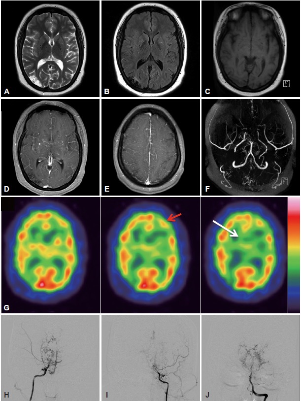
Figure 1.

A, B, and C: Magnetic resonance imaging (MRI) of the brain showed a low signal intensity on the right parieto-occipital area and multiple flow-voids in both basal ganglia on T2-, T1-, and fluid attenuation inversion recovery-weighted images. D and E: Contrast-enhanced MRI showed diffuse leptomeningeal enhancement along the cortical sulci and strong enhancement of perforating arteries in the basal ganglia and deep white matter (“ivy sign”). F: Magnetic resonance cerebral angiography revealed a severe stenosis of both internal carotid arteries at the supraclinoid portion with numerous collateral vessels. G: A 99mTc-hexamethylpropylene amieoxime brain single photon emission computed tomography showed decreased perfusions in the right temporo-occipital cortex and bilateral frontal areas (red arrow) and in both basal ganglia (white arrow). H, I, and J: Digital subtraction cerebral angiography confirmed the moyamoya disease (Suzuki grade IV).
