
Figure 1.
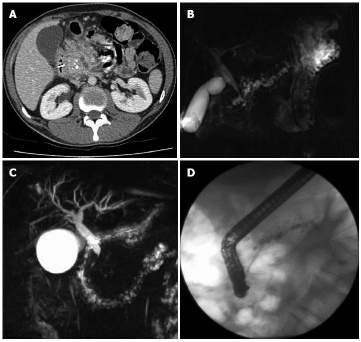
Computed tomography demonstrating enlarged head of pancreas with coarse calcification and a dilated main pancreatic duct (A), magnetic resonance cholangiopancreatography showing a tortuous, dilated pancreatic duct (B), inflammatory stricture of the distal common bile duct (C), endoscopic retrograde cholangiopancreatography showing a stent placed in a dilated pancreatic duct (D).