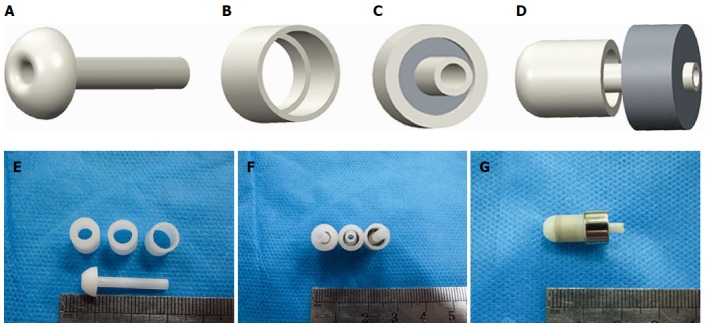
Figure 2.

Mode pattern and real pictures of magnet devices. A: Lateral view of an internal drainage tube; B: View of the biliary part magnet shell; C: Antapical view of the combined biliary part; D: Lateral view of biliary part approach to enteric part; E: The internal drainage tube and shells of different crimping areas; F: Magnets with different pressures; G: Biliary part and enteric part coupled together.
