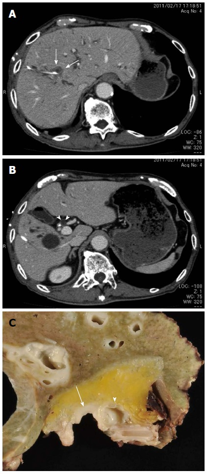
Figure 1.

Preoperative images. A: Enhanced computed tomography showing a 2.2-cm low-density tumor (arrows) in the right hepatic hilum. The white tube is the percutaneous transhepatic biliary drainage tube; B: Enhanced computed tomography showing a cystic lesion (arrowheads) by the common bile duct; the endoscopic nasobiliary drainage tube appears as a white dot. A solitary cyst can also be observed in the right lobe; C: Surgically resected specimen showing a cystic lesion (arrowhead) in the hilar connective tissue near the common bile duct involving the carcinoma (arrow).
