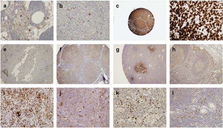
Figure 3.
Lymphoma samples were tested for the expression of Tenth Human Leukocyte Differentiation Antigen (HLDA10) monoclonal antibodies (mAbs) by immunohistochemistry of paraffin sections. (a) mAb to CD1a (10-03) on reactive lymph nodes and (b) lymph nodes involved with Hodgkin lymphoma (HL). (c) mAb to LPAP (10-04) on lymphomas and reactive lymph nodes. (d) LPAP (10-19) did not stain the Reed–Sternberg cells in HL (arrow). (e) The 10-04 mAb did not stain non-lymphoid cells in the liver tissue. (f) LPAP (10-19) weakly stained all lymphoid cells in lymphomas. (g) FAT1 cadherin (10-16) on reactive lymph node specimens. (h) Calreticulin (10-23) on reactive lymph nodes and (i) showing increased staining intensity in Reed–Sternberg cells in HL and (j) scattered large cells in HL and follicular lymphoma (FL). (k) The IL-13-Ra2 (10-37) mAb on HL and (l) on reactive lymph nodes.

