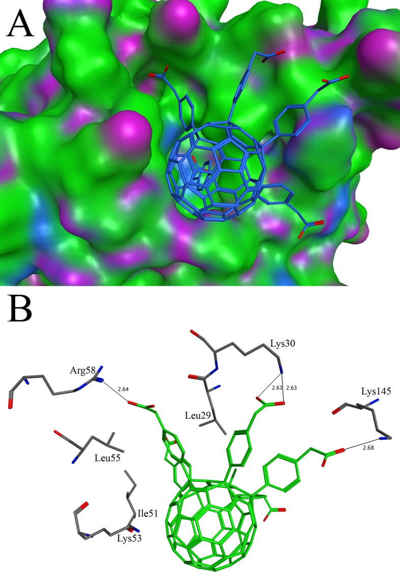
Fig 6. Docking of C60-10 with dihydrofolate reductase: (A) The binding pattern of C60-10 in the active site of dihydrofolate reductase. The C60-10 is shown in stick while dihydrofolate reductase is shown in surface model (B) The diagram of the interactions bewteeen C60-10 and dihydrofolate reductase.

The C60-10 makes interactions with Leu29, Lys30, Ile51, Lys53, Leu55, Arg58 and Lys145. The hydrogen bonds are shown in thin lines.
