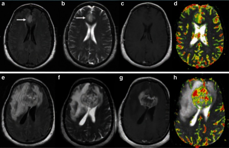
Fig. 3.
MR perfusion images for a pathologically proven mixed oligoastrocytoma. Images a–d show baseline examination (a FLAIR; b T2-weighted pre-contrast; c T1-weighted post-gadopentetate dimeglumine; d dynamic susceptibility-weighted perfusion image with relative CBV color overlay map), which demonstrated a high baseline rCBV of 4.23, indicative of a high-grade tumor. Images e–h show the equivalent sequences at 18-week follow-up, and demonstrate substantial increase in tumor volume and a rCBV of 13.37 [100]. rCBV relative cerebral blood volume, FLAIR fluid-attenuated inversion recovery, MR magnetic resonance. Reproduced from Law et al. [100], Copyright 2014, with permission from The Radiological Society of North America (RSNA®)

