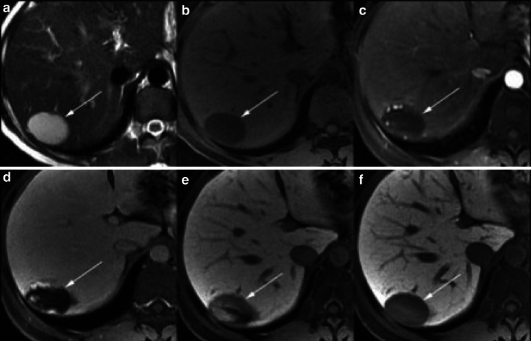
Fig. 4.
Magnetic resonance imaging of the liver in a patient with chronic hepatitis C. Images a and b show pre-contrast T2- and T1-weighted images, respectively; images c–f were obtained 20 s, 1, 5 and 25 min, respectively, after injection of gadoxetic acid. A lesion in segment VII of the liver demonstrates hyperintensity on pre-contrast T2-weighted image, and hypointensity on T1. Post-contrast, the mass has peripheral puddling of contrast in the arterial phase (c) which progressively coalesce (d, e). In the hepatocyte phase (f), the mass is hypointense to the liver with similar signal intensity to blood vessels. Imaging features are characteristic of haemangioma [135]. Reprinted from Cruite et al. [135], with permission from the American Journal of Roentgenology

