
Fig. 11.
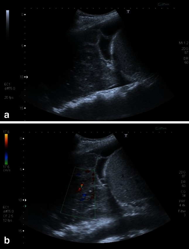
a Sonographic image of a right-sided consolidation. The consolidation shows areas of lower echogenicity suggesting lymphoma infiltration. Additionally a pleural effusion with fibrin strands is present. b Using color-Doppler the hypoechoic areas surround the vasculature (see Online Resources 6 and 7)