
Fig. 4.
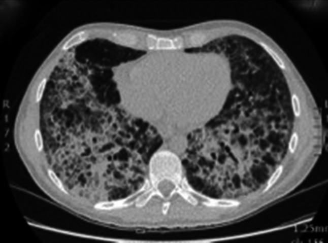
Chest CT after development of respiratory distress showing coarse interstitial thickening with cyst formation and areas of unaffected parenchyma suggestive of pneumocystis pneumonia, which was later microbiologically confirmed in BAL
Official websites use .gov
A
.gov website belongs to an official
government organization in the United States.
Secure .gov websites use HTTPS
A lock (
) or https:// means you've safely
connected to the .gov website. Share sensitive
information only on official, secure websites.
Chest CT after development of respiratory distress showing coarse interstitial thickening with cyst formation and areas of unaffected parenchyma suggestive of pneumocystis pneumonia, which was later microbiologically confirmed in BAL