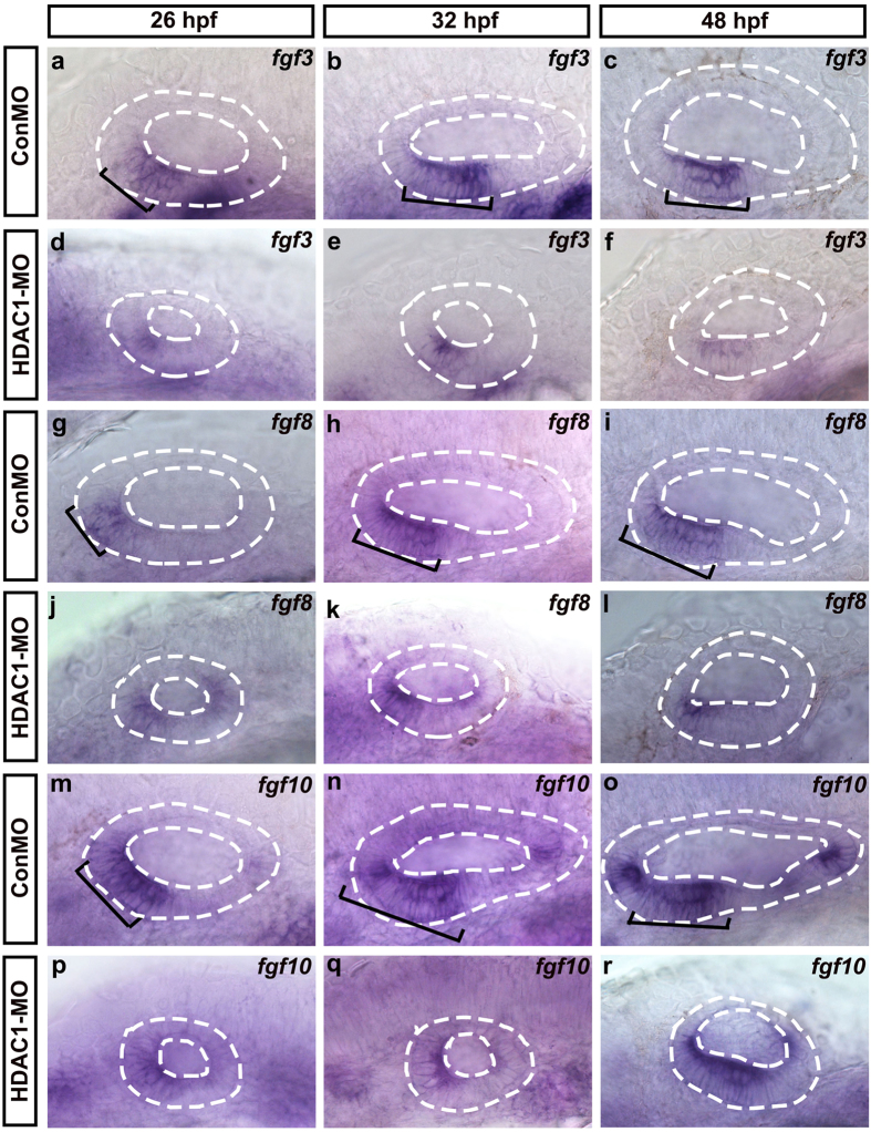
Figure 6. The effect of HDAC1 knockdown on the expression of fgf3/8/10 in the developing inner ear.
Whole-mount in situ hybridizations with fgf3 (a–f), fgf8 (g–l), and fgf10 (m–r) probes in control embryos (a–c,g–i,m–o) and HDAC1 MO-injected embryos (d–f,j–l,p–r) at different developmental stages (as indicated). Black brackets indicate the localization of fgf3-expressing (a–c), fgf8-expressing (g–i), and fgf10-expressing (m–o) cells. Embryos injected with HDAC1 morpholino show reduction in levels of fgf3 (d–f), fgf8 (j–l), and fgf10 (p–r) expression. The otic vesicles are outlined by dashed lines. All images show lateral views with the anterior to the left and the dorsal side up.

