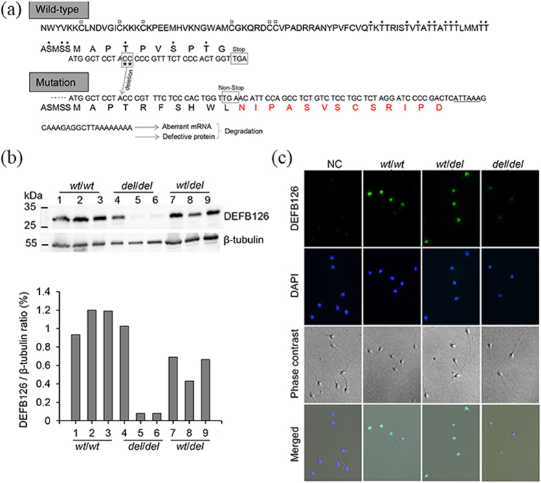
Figure 4. The expression and location of DEFB126 on human sperm.
(a) The wild-type DEFB126 and its two-nucleotide deletion mutant deduced from the mRNA nucleotide sequence; the six highly conserved cysteines (C) marked with empty squares45; seventeen potential residues (serine and threonine, dotted) at carboxyl terminus of DEFB126 predicted to be O-linked glycosylated (NetOGlyc 3.1)43; the two missed nucleotides (CC) labeled with asterisk and its frame-shifted version shown in the lower panel; the additional amino acids in the mutant protein displayed with the red front; the regulatory element of polyA signal sequence labeled with underline. (b) Representative Western blot from the ejaculated sperm of different genotypes (wt/wt, n = 3; wt/del, n = 3; del/del, n = 3), showing unstable expression of DEFB126 in sperm with del/del compared to the sperm with the other two genotypes (wt/wt or wt/del); β-tubulin used as loading control. (c) Localization of DEFB126 (green) on human sperm of different genotypes (wt/wt, wt/del and del/del); the sperm smears stained with polyclonal antibody against DEFB126 (rabbit anti-human β-defensin 126, sc-85535; 1:200 dilution) followed by an Alexa Fluor 488 conjugated donkey anti-rabbit IgG (1:200 dilution); sperm nuclei stained with DAPI (blue); Negative control (NC) sperm staining with rabbit IgG showing no immunoreactive staining.

