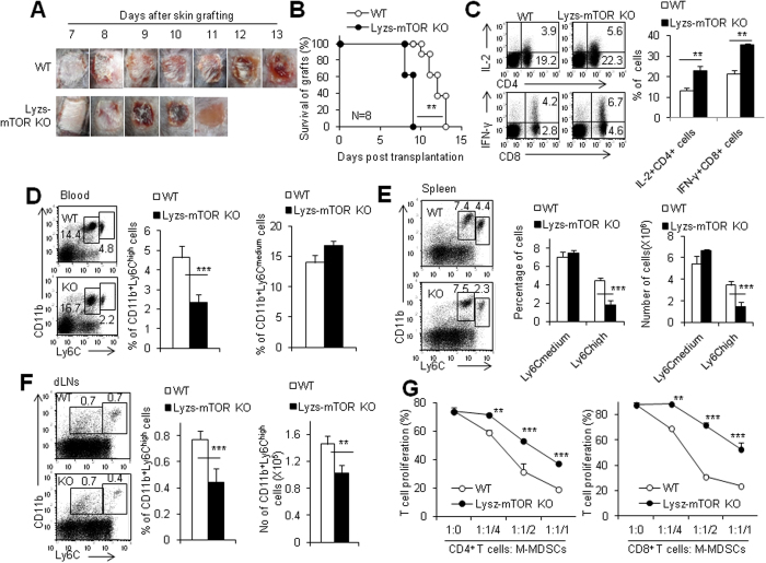
Figure 2. Deletion of mTOR specifically in myeloid cells decreases M-MDSCs in mice after transplantation.
(A) Age and gender matched WT and lyzs-mTOR KO mice were transplanted with allogeneic BALB/c tail skin and macroscopic pictures of alloskin grafts were collected at different time points. (B) Alloskin graft rejection in lyzs-mTOR KO recipients was significantly accelerated compared with WT recipients. The splenocytes, peripheral blood cells and draining lymph node cells of alloskin-grafted WT and Lyzs-MTOR KO recipients were isolated and analyzed by flow cytometry at day 7 after skin grafting. (C) The expression of IFN-γ and IL-2 proteins in gated CD4+ T cells and CD8+ T cells in draining lymph node of WT and lyzs-mTOR KO recipients as determined by intracellular staining. (D) The percentages of CD11b+ Ly6Chi M-MDSCs and CD11b+ Ly6Cmed G-MDSCs in peripheral blood of WT and lyzs-mTOR KO recipients. The percentages and cell numbers of CD11b+ Ly6Chi M-MDSCs and CD11b+ Ly6Cmed G-MDSCs in spleens (E) and dLNs (F) of WT and lyzs-mTOR KO recipients. (G) Isolated CD11b+ Ly6Chi cells from spleens of WT and lyzs-mTOR KO recipients at day 7 after skin grafting were added at different ratios in T cell proliferation system for 72 h. Proliferation was measured by CFSE dye dilution. The numbers in FACS plots represent the percentages of cells in the gates. Results are expressed as mean ± SD. Each group included from 4 to 6 mice and at least three independent experiments showed similar results. **p < 0.01, and ***p < 0.001 compared between the indicated groups.

