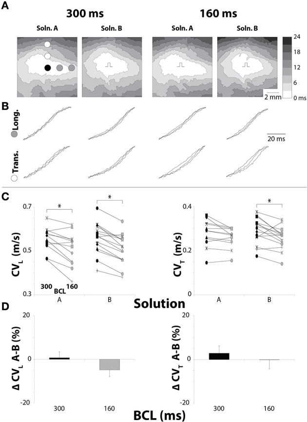
Figure 1.

Pacing rate but not solution composition alters CV in hearts with normal GJ coupling. (A) Representative isochrones from hearts perfused without CBX, showing conduction slowing between pacing rates, but not solutions. (B) Uniformly spaced action potential upstrokes from the same hearts as pictured in (A), demonstrating temporal upstroke separation as another indicator of decreased CV. (C) Black symbols: 300 ms BCL, Gray symbols: 160 ms BCL. CV measurements for longitudinal and transverse directions. CV decreased due to increased pacing rate for each combination except Solution A in the transverse direction. (D) Percent changes in CVL and CVT between Solutions A and B show no changes in CV due to perfusate. *p < 0.025 between pacing rates.
