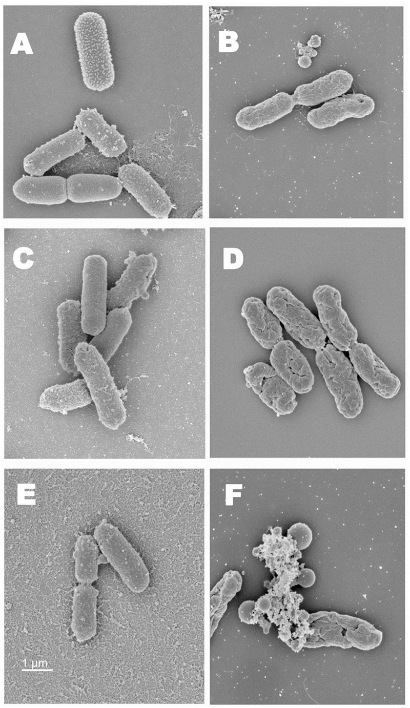
FIGURE 3.

(A) Scanning electron microscopy images of normal EHEC cells (Five-strain cocktail). (B) SEM images of EHEC cells exposed to heat. (C) SEM images of EHEC cells treated with RV (0.2%). (D) SEM images of RV treated EHEC cells exposed to heat. (E) SEM images of EHEC cells treated with RV in CH. (F) SEM images of RV + CH treated bacterial cells exposed to heat.
