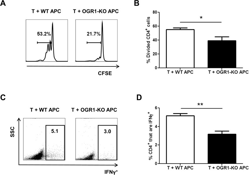
Fig 6. APC from OGR1-KO mice have a decreased ability to support T cell proliferation and cytokine production.
Splenocytes were isolated from WT and OGR1-KO mice and T cells were depleted by MACs separation as described in the Methods. T cell-depleted splenocytes (APC) were stimulated with LPS overnight, washed and co-cultured (1:1 ratio) with CFSE-labeled WT CD4+ T cells that were plated onto anti-CD3 antibody-coated plates. The percentage of divided CD4+ cells (A-B) was examined after 3 days of culture. Data shown are from 4 individual experiments. (C-D) Cells were stimulated with PMA, ionomycin in the presence of brefeldin A for an additional 4 hours and intracellular staining for IFN-γ was performed. (C) shows representative FACs plots of IFN-γ expression versus side scatter of CD4+ cells and (D) shows the means + SEM of values obtained from 3 individual experiments. *p<0.05, **p<0.01 by t-test (two-tailed).

