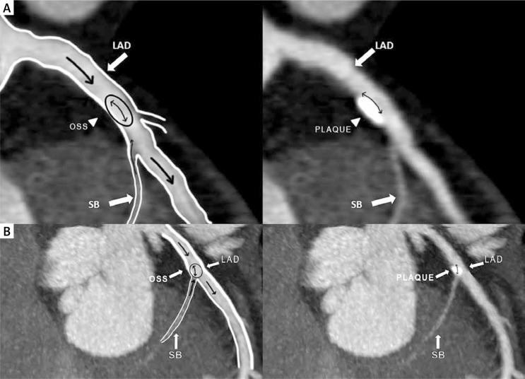
Fig. 1.
Panel A and B. Multiplanar reconstructed images of the left anterior descending artery (LAD) and the septal branch. Diagram illustrating plaque location in the proximal segment of the LAD artery. Note the anatomy of the septal branch and its intramyocardial course. In proximity to the septal branch ostium, the LAD endothelial cells are exposed to atherogenic low oscillatory shear stress, which leads to plaque formation (left panel). On the right, the corresponding region of the plaque location (myocardial bridging effect)
LAD – left anterior descending artery, OSS – oscillatory shear stress, SB – septal branch

