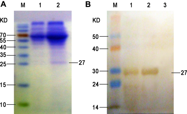
FIGURE 1.
SDS-PAGE (A) and Western blot (B) of the expressed ompA in the supernatant. (A) M, protein molecular weight markers; lane 1, proteins isolated from the culture supernatant of BHK-21 cells at 72 h after empty plasmid transfection (negative control); lane 2, proteins isolated from the culture supernatant of BHK-21 cells at 72 h after pVAX1-ompA plasmid transfection. (B) M, PageRuler prestained protein ladder; 1, proteins isolated from the culture supernatant of BHK-21 cells at 72 h after pVAX1-ompA plasmid transfection; 2, purified recombinant protein from eluted fraction; 3, proteins isolated from the culture supernatant of BHK-21 cells at 72 h after empty plasmid transfection (negative control).

