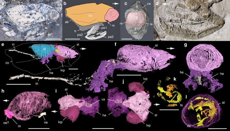
Figure 1. External and internal morphology of Dollocaris ingens.
Thylacocephala; Middle Jurassic La Voulte Lagerstätte. (a,b) MNHN.F.R50939, right lateral view. (c,h) MHNL-20293244, frontal view showing the pair of bulbous eyes and details of anterior part of digestive system in lateral view. (d) MNHN.F.R06202, details of prehensile appendages. (e–g,k,l) UJF-ID-1799, XTM reconstructions of internal anatomy (respiratory and digestive system), general view and details of digestive system in lateral and anterior views, and crustacean exoskletal fragments inside the stomach. (i,j) FSL 710067, XTM reconstructions of the anterior part of digestive system in lateral and dorsal views. White arrows indicate front part. ab, abdomen; ca, carapace; cl, claw; co, carapace outline; da, dactylus; gi, gills; he, hepatopancreas; in, intestine; le, left eye; lvp, latero-ventral pouch; m, mouth; oe?, possible oesophagus; pa1-3, 1st to 3rd pair of prehensile appendages; re, right eye; st, stomach; tr, trunk; ts, trunk sclerite (fragment); va, valve. Scale bars, 10 mm.

